What do the tests before surgery mean?
A number of tests are performed before surgery and some tests may need to be repeated. Tests may be done for different reasons, such as to find the diagnosis or to find out how advanced a cancer is. Before surgery we do tests to check your fitness and for unexpected problems. Some tests help us plan how to do the operation. Some results will be available immediately but most tests will take several days for the results.
After a test you should have a follow up appointment booked or we may phone to speed things up. If you are not sure what will happen next, or if you have extra questions please ask your doctor or nurse.
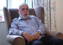
Martin’s story – Symptoms and diagnosis
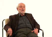
David’s story – Symptoms and diagnosis
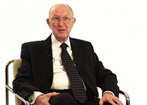
Charles’ story – Symptoms and diagnosis
Flexible bronchoscopy
A bronchoscopy (bron-kos-copy) is a common test used to examine the lungs and airways, it is done using local anaesthetic (you are awake but can have the throat numbed or some sedative drugs). The doctor passes a thin flexible camera through your nose or mouth, through the windpipe (trachea) and into the lungs. Photographs and samples can be taken. Samples are then looked at under a microscope to try and find the diagnosis.
CT scan
CT scans use X rays to take picture of the inside of the body from different angles. The pictures are combined to build a detailed picture of the inside of the body. This gives more information than one X ray on its own. The scan is painless and takes around 10 minutes, an injection may be needed.
ECG (electrocardiogram)
This is commonly called a heart tracing. 12 stickers are put on the chest and connected to leads. The electrical activity in the heart is measured and printed out. This allows doctors to assess the heart rhythm. The tracing is painless, taking the stickers off may be uncomfortable if you have hair on your chest.
Echo (echocardiogram)
An echo is an ultrasound scan to examine your heart. Jelly and a probe are placed on the skin in the same way pregnant women have scans. The test is not painful but the operator may need to press on the skin. It may be needed if you have had heart problems or breathing problems.
Lung function tests
These tests show the doctor how well your lungs work. You blow into machines via a mouthpiece and they record how much air you can breathe in or out and how much air (oxygen) your lungs absorb. The results help decide on appropriate treatment for you by predicting how your body would cope with part of a lung removed.
Exercise tests
Exercise tests measure your overall fitness. They are sometimes needed if other tests show you may have a problem with your heart or your breathing. The tests may be done in the various ways, such as measuring how far you can walk or exercising on a bike.
Perfusion or ventilation scan
This is a simple test to check the function of different parts of the lung. A perfusion scan measures blood flow to the lung, a ventilation scan measures airflow to the lung. An injection of radioactive fluid is needed for the scan.
PET scan
A PET scan uses small amounts of a radioactive sugar to check how active cells in the body are. Cells may be more active for a number of reasons, including infection or cancer. A PET scan cannot tell us why cells are more active. Looking at the PET scan and the CT scan gives important information to plan the best treatment. PET scans are used to assess the stage of lung cancer.
Blood tests
Before surgery we check the function of your kidneys and liver, blood clotting and blood type. We check for anaemia (your haemoglobin level), the levels of white blood cells and platelets. These can usually all be done from taking blood samples from one needle.
You will have a blood test the first day after your operation and again if we need to monitor one of the tests.
MRI scan
MRI scans make pictures using magnets, not radiation. Water and fat in the body look different on MRI scans. They are very useful for assessing lumps near the spine. The scan involves lying in a tube, if you panic in small spaces please tell your doctor.
Biopsies
A biopsy means taking a small piece of tissue (sample) from the body and sending it for analysis in the laboratory. This involves looking at the tissue structure under a microscope. It helps us diagnose the cause of certain lumps in the body. There are different ways of taking a biopsy. Sometimes the biopsy doesn’t tell us the diagnosis. This may be because the piece of tissue was too small or there was too much scar tissue.
An endobronchial ultrasound (EBUS) is like a bronchoscopy and done using local anaesthetic and sedation. The flexible camera has an ultrasound probe attached which allows the doctor to see any enlarged glands (lymph nodes) through the wall of the wind pipe. Samples of the glands can then be taken. Samples are then looked at under a microscope to try and find the diagnosis.
A CT scan may show a shadow that is easy to get to using a needle to get a sample (biopsy). A radiology doctor will use the CT scan to decide the best place to take a sample from using the CT scanner. The doctor does an injection to numb part of the chest (local anaesthetic) then takes the sample with a needle. The sample is looked at under the microscope to find the diagnosis. Most people do not have a problem having the biopsy, other people have discomfort. If you are in pain or want to stop please tell the doctor and they can help you.
Surgery may be recommended both to get a biopsy and help treat symptoms.
The following are operations that may be done to get a diagnosis:
- Biopsy by a cut on the neck (cervical mediastinoscopy)
- Biopsy by a cut on the front of the chest (anterior mediastinotomy)
- Pleural biopsy
- Lung biopsy
- Bronchoscopy
- Frozen section
Lung tissue and lymph glands which are removed are sent to the laboratory to be looked at under the microscope, this is called histology. It is a delicate process and takes about 2 weeks to get results. Histology can help us diagnose lung disease and also assess the stage of lung cancer. Histology results will be discussed with you in clinic.